 Other names:
1. Adenoameloblastoma
2. Ameloblastic Adenomatoid Tumour
Definition:
It is a benign tumour of odontogenic origin mostly associated with unerupted or impacted maxillary cuspids or canines i.e tooth bearing areas of jaws and resembles cytologically too the dental lamina and components of enamel organ i.e. odontogenic origin.
Note:
- Some people says it a benign neoplasm, while other says it is a hamartomatous malformation.
- It is called 2/3rd tumour because 2/3rd cases present in
1. Maxilla
2. Female
3. Canine
4. Unerupted tooth
Histogenesis:
Like all other odontogenic tumours, specific stimulus that triggers proliferation of progenitor cells of AOT is unknown. It is odontogenic in origin. AOT can arise from the
2. The epithelial lining of dentigerous cyst
3. Epithelial rests of Malassez of the tooth
4. Remnants of dental lamina
Clinical features:
Age: <20 years, F>M, Maxilla>Mandible,
Site: Anterior r jaw> Posterior jaw (Cuspids, most common)
- Asymptomatic but swelling of size 1.5 to 3 cm with facial asymmetry
- If extraosseous (in gingiva): Painless, gingival coloured mass
(Maxillary gingiva> Mandibular gingiva, 10 times
Differential diagnosis:
1. Ameloblastoma
2. Gingival fibroma
3. Peripheral cemento-ossifying fibromas
4. Calcifying odontogenic cysts and tumours
Radiographic features:
- Unilocular radiolucency with smooth corticated (sometimes sclerotic) border usually pericoronal or juxta coronal or sometimes extended beyond CEJ
- Radiopaque foci (in 65% cases) within radiolucent lesion, eccentrically placed
- Divergence of root, resorption of roots and displacement of teeth
- Erosion of underlying alveolar bone cortex
Histologic features:
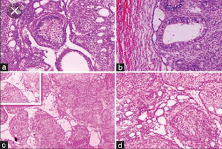
- Multinodular proliferation of spindle, cuboidal and columnar cells forming nests or rosette-like structures
- Scattered duct like structure with lumen of varying size, lined by single layer of cuboidal to columnar epithelium (nucleus away from lumen)
- Lumen are lined by an eosinophilic ring called hyaline ring
- Between cell-rich nodules, stellate reticulum like spindle cells with small amount of calcifications
- Liesegang rings can be seen in stroma (Irregular to round calcified bodies which exhibit areas with a concentric layered pattern)
- Stroma: loose, hypocellular and fibrovascular
Treatment: Conservative surgical excision
Prognosis: Good
MCQS From Adenomatoid Odontogenic Tumour: Click Here


No comments:
Post a Comment